How are leading research institutions working with label-free live cell imaging in 2026? At the start of April, scientists from MIT, Mass General Hospital and Harvard Medical School shared their research using Nanolive imaging to study cell death and CAR T engineering. This newsletter edition recaps some highlights from the seminar and announces Nanolive’s latest data walkthrough for liquid tumor analysis. Finally, we highlight April 2026’s key publications featuring label-free live cell imaging and AI analysis in other fields including aging, drug discovery, and stem cell differentiation.
Ferroptosis investigation at the population and sub-cellular levels
Capturing propagation of cellular phenomena at the population level requires live cell imaging, and noninvasive imaging allows data capture that is unbiased by fluorescence, which is particularly important when studying cell death. In this seminar, Ryan Mansell from the Koch Institute for Integrative Cancer Research at MIT presented his work quantifying ferroptotic cell death label-free using Nanolive’s LIVE Cytotoxicity Assay. His timelapse imaging of cells at the population level revealed a ‘ferroptotic wave’ phenomenon that illustrates how ferroptosis propagates through cell-cell contact.
Using deeper analysis at the subcellular level using Nanolive’s organelle-based assays, Ryan also revealed phenotypic changes in mitochondria and lipid droplets which correlated with ferroptosis progression.

Figure from Ryan Mansell’s talk “A New Window into Ferroptosis: Label-Free Imaging of Cellular Death” showing population-level timelapse imaging of cells undergoing ferroptosis. The digital overlay shows living cells colored green, and ferroptotic cells colored blue.
CAR-T engineering
The dynamics of CAR-T activity are equally suited to label-free live cell imaging. Dr Sangwoo Park from Mass. General Hospital and Harvard Medical School presented his research titled “Engineering Immune Cells to Overcome the Cancer Cell Glycocalyx”. After engineering CAR-T cells to better target cancer cells, Dr Park used Nanolive’s label-free LIVE T Cell Assay to monitor the dynamics of CAR-T expansion and killing of cancer cells.

Figure from Dr Sangwoo Park’s talk “Engineering Immune Cells to Overcome the Cancer Cell Glycocalyx”. The digital overlay shows target cancer cells colored blue, CAR-T cells colored red, and areas of interaction colored yellow.
Watch the seminar on demand
The full seminar including researcher talks, Q&A, and introduction to live cell imaging by Dr Joanna Sadowska are available on demand.
You can watch it here.

Explore the Nanolive workflow for cell therapy assessment

From activation to target engagement and killing, Nanolive’s revolutionary technology enables the identification of the most effective therapeutic candidates using live, label-free and automated analysis of effector cell activation, interactions, and killing. This data walkthrough presents insights from nonadherent AML and CML models with gamma-delta T cells, and human CAR T cells, using the only high-resolution and automated system for quantifying the effector cell response in co-culture.
Watch the recording on demand here.
Latest publications
- Stem cell differentiation: Alsolami, S. et al., (2026) ‘A single small molecule-based human embryo model reveals V-ATPase requirement in mammalian blastocyst cavitation’, Cell Research, https://doi.org/10.1152/ajpendo.00470.2025.
- Aging: Saha, S. et al., (2026) ‘Holotomography-based, label-free quantification of cellular dry mass as a biophysical indicator of microglial Aβ phagocytosis during senescence’, Biogerontology, https://doi.org/10.1007/s10522-026-10421-4.
- Aging: Singam, A. et al., (2026) ‘Unveiling Lipid Droplet Transport Dynamics as Biomarkers of Senescence Using Label-Free, Time-Lapse Holotomography’, Aging and Disease, http://dx.doi.org/10.14336/AD.2024.1408.
- Cell death: Brahim, S. et al., (2026) ‘Kremen1 dependence receptor induces SEC24C and ATG9A-dependent cell death’, Cell Communication and Signalling, https://doi.org/10.1186/s12964-026-02790-7.
- Drug discovery: Rusak, A. et al., (2026) ‘The CHI3L1 protein plays role as a protecting factor against autophagy in glioblastoma cells’, Apoptosis, https://doi.org/10.1007/s10495-026-02319-w.
- Drug discovery: Castillo, U. G. et al., (2026) ‘Diels–Alder Adducts from Maytenus chiapensis’, IJMS, https://doi.org/10.3390/ijms27073318.
- Immunology: Alsolai, S. et al., (2026) ‘Stable bioreactor control reveals acidic pH–driven metabolic reprogramming and mitochondrial dysfunction in human lymphoblastoid cells’, Communications Biology, https://doi.org/10.1038/s42003-026-10013-5.
You can find over 400 publications featuring Nanolive imaging here.
Subscribe to this monthly newsletter to stay up to date with AI applications in cell biology by clicking the button at the bottom of the page.
Read our latest news
Move beyond endpoints with dynamic EC50
Online walkthrough – Replace fragmented assays: one workflow for cell therapy assessment
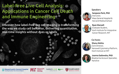
Seminar – Label-Free Live Cell Analysis: Applications in Cancer Cell Death and Immune Engineering
