Nano News
Looking inside Nanolive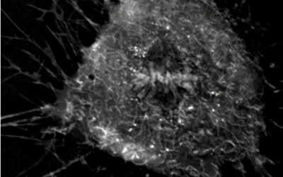
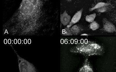
Nanolive imaging captures discrete phases of mitosis in spectacular detail
Nanolive’s label-free, non-invasive imaging allows us access to the dynamics of biological processes, such as mitosis in living cells, with unprecedented resolution.Here, we observe cell division in human mesenchymal stem cells using Nanolive’s 3D Cell Explorer. One...
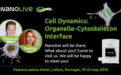
Nanolive at Cell Dynamics: Organelle-Cytoskeleton Interface – Lisbon, Portugal
Nanolive will be at Cell Dynamics: Organelle-Cytoskeleton Interface from 19 - 22 May 2019! We hope to see you there and discuss about new possibilities for label-free live cell imaging in 3D! Meet us at Pestana Palace Hotel, Lisbon, Portugal! Read our latest...

New Publication from The University of Sydney & University of New South Wales
Impact of food additive Titanium Dioxide E171 on human health The University of Sydney & University of New South Wales have recently published their work on the "Impact of the Food Additive Titanium Dioxide (E171) on Gut Microbiota-Host Interaction". They have...
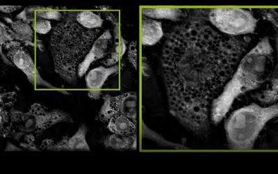
Cell drinking: a closer look on macropinocytosis
Vesicle Transport Vesicle transport is an active and thus energy consuming process that the cell uses in order to capture or release macromolecules into or out of the cell. While exocytosis helps release content like proteins, waste products or toxins to the outside...
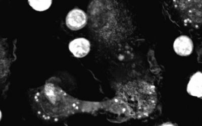
Image of the Day from The Scientist: Macrophages in Action
Image of the day (April 5) of The Scientist! Thanks for featuring a video of these beautiful macrophages absorbing e.coli bacteria! Great video from Michiel van der Vaart from Leiden University! Congrats!Read our latest newsNanolive microscopes Get a quote >3D CELL...
Contest: Nanolive’s Egg Hunt
Easter is around the corner and Nanolive wants you to have some fun! We have hidden plenty of cute Nanolive easter eggs on different pages of our website and in our news section of 2019. One of the eggs is a special one, and we want you to find it! If you find the...
Summer School 2019 in “Practical Holotomographic Microscopy for Live Cell Imaging”
Nanolive is offering for a second time a summer school in “Practical Holotomographic Microscopy for Live Cell Imaging” on the sunny shores of Lake Geneva in August 2019. Join us and experience how you can use this powerful technology to interrogate your...

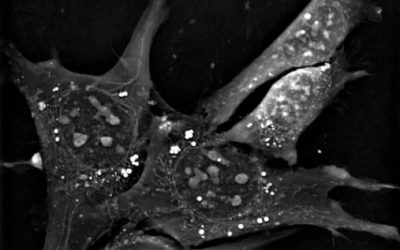
Wildtype vs. Mutant Plasmatocytes in Drosophila Melanogaster
Drosophila melanogaster, the fruit fly, is a model organism widely used in biomedical research thanks to its versatility. Some advantages over other models are its low cost, easy manipulation, short life-cycle and large range of easily performed genetic modifications....

Participate in the Nikon Small World Competition with your Nanolive live cell images & videos!
Nanolive would like to invite you to participate in the Nikon's Small World Competition 2019 with your Nanolive live cell images & videos. This is the perfect place to showcase your beautiful label-free live cell images and videos to a broad global...
New Distribution Partner for the Nordics: RAMCON A/S
Nanolive is proud to announce a new distribution partner for the Nordic countries: RAMCON A/S. RAMCON was established in 1988, and is today a modern company with more than 50 employees in sales, support and service. They focus on these three core...
New Distribution Partner for Canada: Quorum Technologies Inc.
We are happy to announce our new partner in Canada: Quorum Technologies Inc.. Quorum Technologies Inc. is a private Canadian owned enterprise located in Guelph Ontario. The company develops and integrates scientific instruments and is the leading provider of Live...
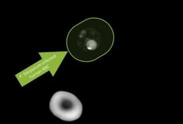
P. falciparum´s intra-erythrocytic cycle, an important immune escape strategy in Malaria
IntroductionMalaria is a preventable and curable disease caused by the genus Plasmodium. In 2022, around 249 million cases were estimated, from which 608000 resulted in death. As many as 85 different countries registered malaria cases. The most severe form of human...
Be good to your skin. You’ll wear it every day for the rest of your life.*
*Quote by Renée Rouleau The skin is the largest organ of the body, spanning an area of 1.8 square meters and forming the border between the body’s inner organs, bones, muscles and the outer environment. Nerve endings and receptors in the skin allow us to interact...
Turn your cell phone into a Cell Phone
Did you know that you can turn your cell phone into a Cell Phone now? Download any of these videos and set them as your wallpaper! Just click on the any of the images below and you will be forwarded to a page to download the video. The links are only working for the...
New Application Note: When Holotomography Meets Immuno-Oncology
In this application note we will discuss new research possibilities that arise from combining holotomography and immuno-oncology, which is more than ever in the spotlight. In fact, the 2018 Nobel Prize for Medicine went to Allison and Honjo for the discovery of a new...
Cell death: 4 ways to die!
This video shows the versatility of the 3D Cell Explorer microscope in imaging both very fast and very slow processes without compromising with cell health and at the desired frequency. There are 4 different cell types. Can you guess the name of each cell death when...
A microscopic view of label-free and living Listeria bacteria
What is Listeria and where does it come from? Infection from Listeria monocytogenes is a food borne bacterial illness that can be very serious for pregnant women and people with impaired immune systems. Listeria infection can be contracted by eating badly preserved...
Label-free live cell imaging of antigen mediated mast cell degranulation in collaboration with the Max-Planck-Institute
In this video you can observe antigen mediated mast cell degranulation. Mast cells reside in connective and mucosal tissues near host-environment barriers throughout the body as a part of the immune system. They serve as immunologic sentinels and are major...