Nanolive will be attending together with its regional partners AXT this year’s ISSCR in Melbourne, Australia from June 20 to 23. We are bringing special prizes to the global stem cell event for anyone who comes to our booth (#132) with the “secret”...
Nanolive is very happy to announce that we have joined the Scale Up Vaud initiative, a program that supports scale-ups companies in their growth phase. We are very proud to be a member of this community since today. This is a great recognition for the efforts of...
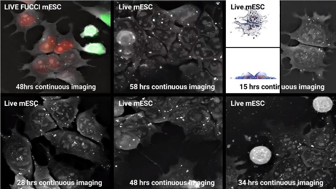
Live FUCCI mESC were imaged for over 48 hours with Nanolive’s 3D Cell Explorer-fluo. A holotomographic image was taken every 14 seconds while a double channel (green and red) epifluorescence image was only taken every 30 holotomographic frames...
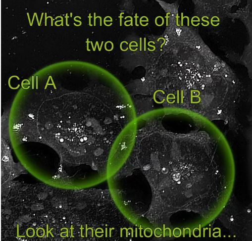
As mentioned before in our dedicated stem cell blog post https://vkv7flpfqbb.c.updraftclone.com/labelfree-stemcells/, studying stem cells is among, if not THE biggest challenge in today’s biological research. Scientists can discover more about the behavior of...
It is almost impossible to image the inside of a living cell without damaging it, even with the most modern devices. Traditional microscopy techniques require researchers to prepare their samples well in advance (1–72 hours). These procedures are likely to be...