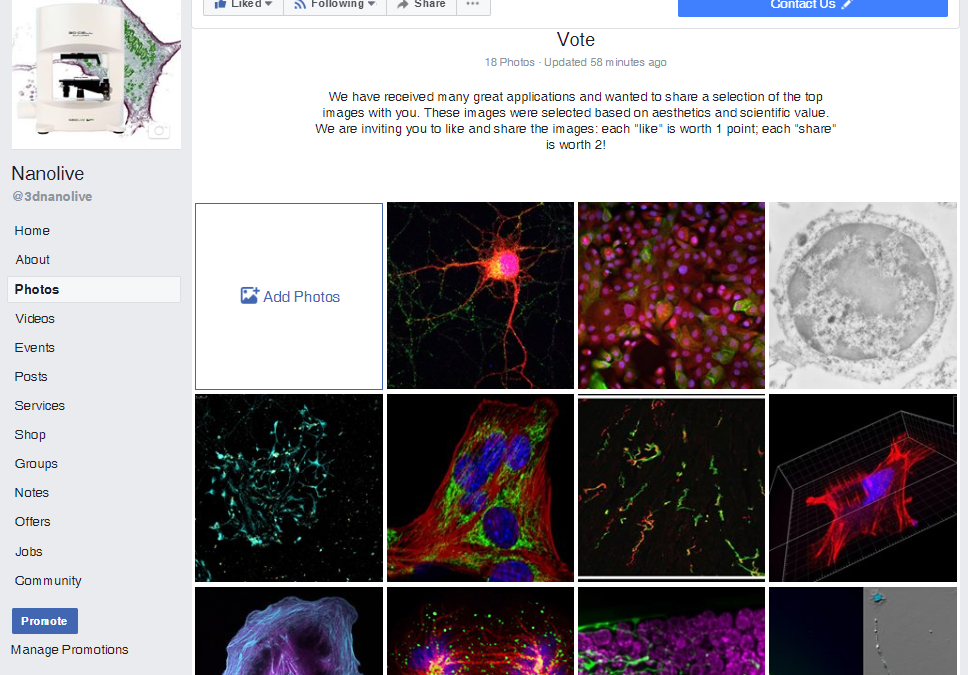
We have received many great applications and wanted to share a selection of the top images with you. These images were selected based on aesthetics and scientific value. As we have many great applications, we would like to put the images to public vote. The winner...
Watch the video and impress us with your live cell imaging knowledge. We are asking you to describe all dynamic processes that you can observe. E-mail us your answer to lookinginsidelife@nanolive.ch and mention the following: Time point and description of...
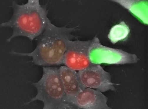
Live FUCCI mESC were imaged for over 48 hours with Nanolive’s 3D Cell Explorer-fluo. A holotomographic image was taken every 14 seconds while a double channel (green and red) epifluorescence image was only taken every 30 holotomographic frames...
Nanolive is proud to announce a new publication in ASC Publications from our users in the University of New South Wales Australia & the University of Paris. Bacterial biofilms are usually difficult to treat and are the major cause of chronic and...
Long Term Live Imaging of Mouse Embryonic Stem Cells Nanolive is offering a summer school in “Practical Holotomographic Microscopy for Live Cell Imaging” on the sunny shores of Lake Geneva in August. Join us and experience how you can use this powerful...