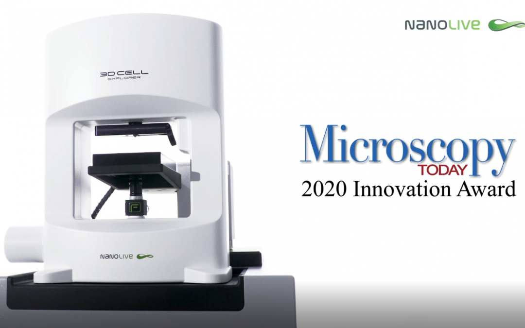
Nanolive are delighted to have been selected as winners of the 11th annual Microscopy Today Innovation Awards in 2020 for the development of our automated microscope, the CX-A, a non-invasive live cell imaging method for continuous organelle monitoring in cell...
We are happy to announce that our webinar with the topic “Unlocking the mysteries of neurite growth in primary cortical neurones: a quantitative approach to live cell imaging” is now available on demand. In this webinar, Dr. Emma Gibbin, Communications...
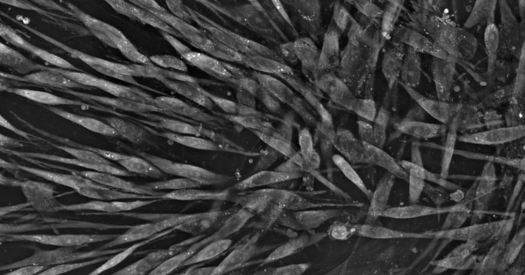
Mesenchymal stem cells (MSCs) hold great promise for regenerative medicines such as cell therapy and tissue engineering (1,2). These approaches require cells to be grown in culture and differentiated into specific cell types such as osteoblasts (bone cells),...
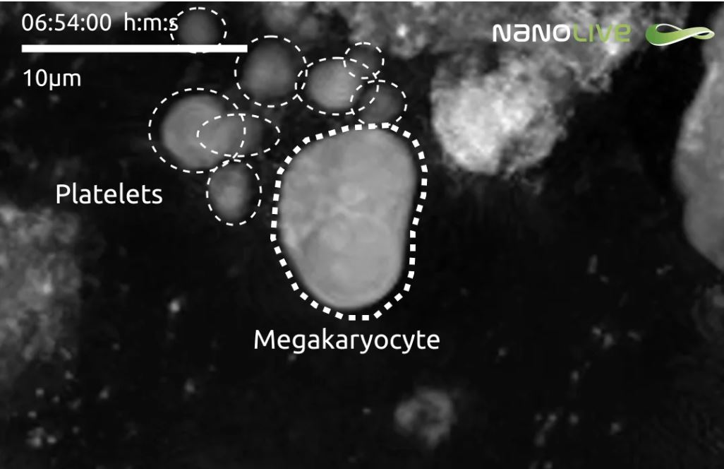
Stem cells were isolated from the umbilical cord matrix and grown in culture for 14 days. At this stage most cells in the population are at the mature megakaryocyte stage and can start producing platelets in internal membranes present in their cytoplasm. In this...
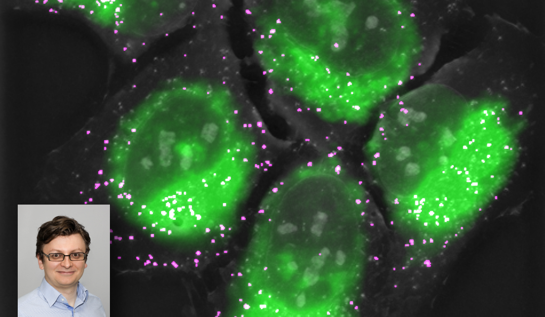
Dr. Michal Cifra is the young, dynamic team leader of the Bioelectrodynamics team at the Institute of Photonics and Electronics Academy of Sciences (Czech Republic). His lab has recently purchased Nanolive’s 3D Cell Explorer-fluo microscope and published his first...