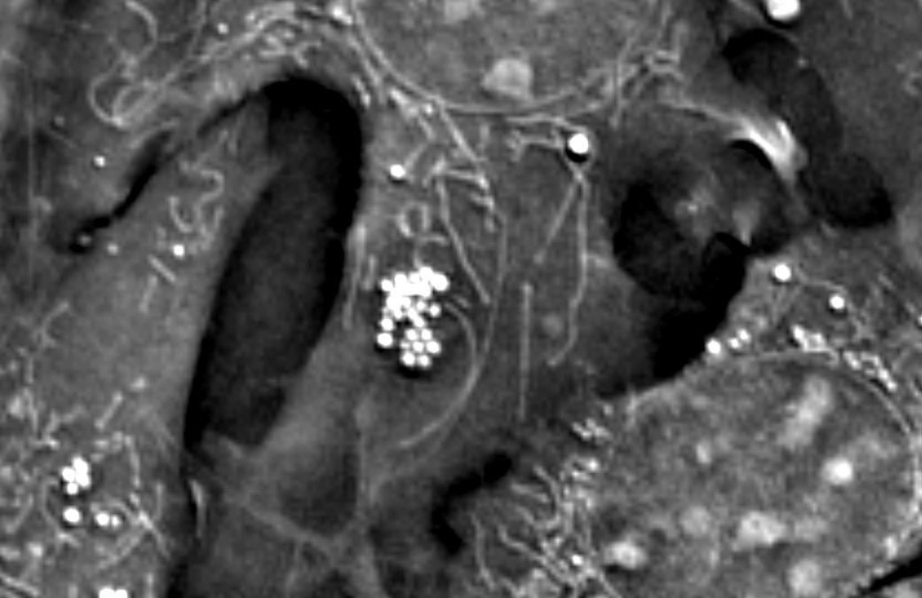
Cellular metabolism is a complex, integrated process that is supported by key organelles, such as lipid droplets and mitochondria, which engage in numerous energetic and signaling mechanisms. A better understanding of the dynamics and interactions of such organelles...
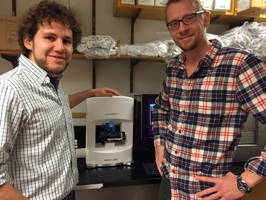
Nanolive is proud to announce the newest addition to our Science Advisory Board: Clemens Grassberger, PhD, of Harvard Medical School. Dr. Clemens Grassberger was Nanolive’s first customer. He marked a historical moment for the live cell imaging company and...
It is almost impossible to image the inside of a living cell without damaging it, even with the most modern devices. Traditional microscopy techniques require researchers to prepare their samples well in advance (1–72 hours). These procedures are likely to...
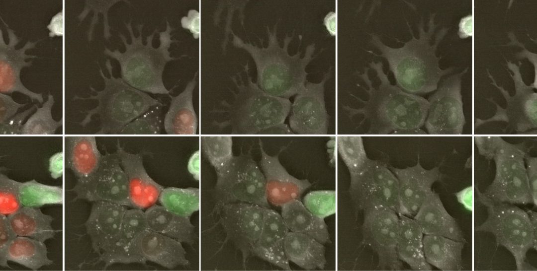
In this application note we will present the time lapse imaging of mouse embryonic stem cells (mESCs) that have been genetically modified to express the fluorescence ubiquitination cell cycle indicator (FUCCI), a two-color (red and green) indicator that allows to...
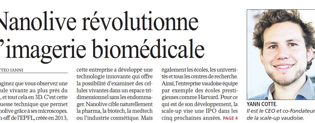
The first featured article as scale-up has been published this morning in the newspaper Agefi. “Until now, it wasn’t possible to observe a living cell and its organelles in 3D without harming it.” Read the full article in French...