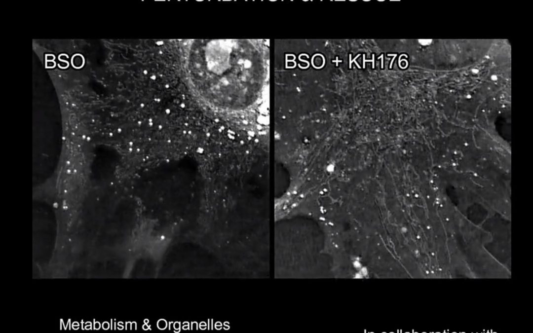
A major problem with current imaging techniques is phototoxicity that leads to the observation of perturbed dynamics. However, the 3D Cell Explorer overcomes this problematic as it injects in the sample ~100 times less energy (~0.2 nW/µm2) than light sheet microscopes...
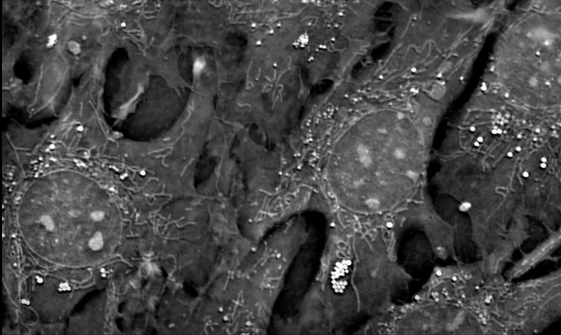
Watch the webinar here! Please register here to view the webinar on-demand Register Dr. Mathieu Frechin, Head of Quantitative Biology at Nanolive introduces you to the advantages of our holotomographic microscope for imaging key organelles supporting cellular...
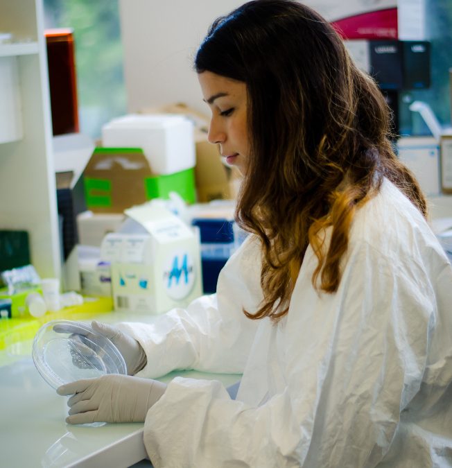
As announced a couple of days ago, Nanolive chose Ms. Valentina Palacio Castaneda as the winner of the “Scientist of Tomorrow” contest. We were keen to know more about the winner and her passion for biology and asked a few questions before her...
It is almost impossible to image the inside of a living cell without damaging it, even with the most modern devices. Traditional microscopy techniques require researchers to prepare their samples well in advance (1–72 hours). These procedures are likely to...
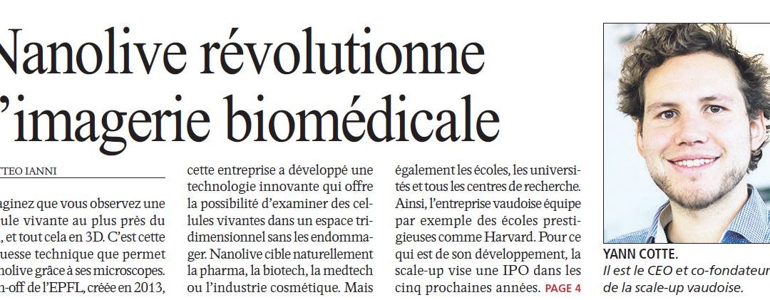
The first featured article as scale-up has been published this morning in the newspaper Agefi. “Until now, it wasn’t possible to observe a living cell and its organelles in 3D without harming it.” Read the full article in French...